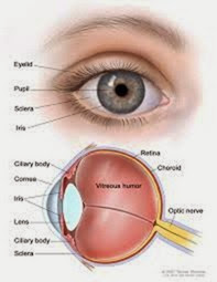
intraocular pressure The pressure within the EYE that maintains the eye’s form and structure. Nor- mal intraocular pressure in an adult is 12 to 22 millimeters of mercury (mm Hg). A device called a tonometer measures intraocular pressure, either through light contact against the anesthetized eye or via the force of resistance to a puff of air blown against the eye’s surface (a noncontact method that does not require anesthetic drops). Elevated intraocular pressure is called ocular hypertension. Lower than normal intraocular pressure is called ocular hypotension.
Ocular hypertension (greater than 21 mm Hg) is a hallmark sign of GLAUCOMA, an eye condition that, if untreated, results in complete loss of vision. Other health conditions that can increase intraocular pressure include tumors that press against the eye, ORBITAL CELLULITIS, and GRAVES’S OPHTHALMOPATHY. Increased intraocular pressure damages the cells on the front of the OPTIC NERVE (the retinal ganglia), leading to permanent VISION IMPAIRMENT. Ophthalmic medications that reduce intraocular pressure work through various mechanisms, depending on the cause of the increased pressure.
Ocular hypotension, in which the intraocular pressure is lower than normal (less than 12 mm Hg), characterizes chronic UVEITIS (INFLAMMATION of the structures of the eye) and of certain tumors of the eye. Ocular hypotension also sometimes accompanies systemic HYPOTENSION (low BLOOD PRESSURE) and as a SIDE EFFECT of medications, notably general anesthesia agents.
See also OPHTHALMIC EXAMINATION; TONOMETRY; VITRECTOMY; VITREOUS DETACHMENT.
iritis INFLAMMATION of the iris, the MUSCLE surrounding the pupil of the EYE. Iritis may develop
as a result of INFECTION, such as CONJUNCTIVITIS that spreads to involve other structures of the eye. Iritis also occurs as part of the inflammatory process in AUTOIMMUNE DISORDERS such as RHEUMATOID
ARTHRITIS. The symptoms of iritis include
• irritation and a “gritty” sensation in the eye
• redness of the eye
• PHOTOPHOBIA (sensitivity to bright light)
• blurry vision
• excessive tearing
The ophthalmologist can diagnose iritis based on the appearance of the iris and the eye, though will additionally perform a SLIT LAMP EXAMINATION to look for involvement of other structures of the eye. Treatment is ophthalmic ANTIBIOTIC MEDICA- TIONS (eye drops or ointment) when the ophthalmologist suspects bacterial infection and ophthalmic CORTICOSTEROID MEDICATIONS to reduce the inflammation. When the cause of the inflammation is systemic, the ophthalmologist may pre- scribe anti-inflammatory medications such as corticosteroids or NONSTEROIDAL ANTI-INFLAMMATORY DRUGS (NSAIDS). Treatment usually resolves the symptoms without residual VISION IMPAIRMENT. Untreated or recurrent iritis can have long-lasting effects on vision, including increased INTRAOCULAR PRESSURE.
See also BACTERIA; EPISCLERITIS; GLAUCOMA; KER- ATITIS; SCLERITIS; UVEITIS.
ischemic optic neuropathy Damage to the OPTIC NERVE resulting from insufficient blood supply, sometimes called “STROKE” of the optic nerve. Ischemic optic neuropathy is occurs most commonly in people over age 55 and causes mild to complete VISION IMPAIRMENT. There are two forms: arteritic, associated with giant-cell arteritis (an inflammatory disorder of the arteries that typically affects the temporal arteries) and nonarteritic, which correlates with CARDIOVASCULAR DISEASE (CVD) such as HYPERTENSION (high BLOOD PRESSURE) and ATHEROSCLEROSIS. Other conditions associated with the nonarteritic form include DIABETES, HYPOTENSION (low blood pressure), and RHEUMATOID ARTHRITIS.
Vision impairment due to ischemic optic neuropathy comes on suddenly. In a characteristic pat- tern, a person wakes up in the morning with noticeable loss of VISUAL ACUITY and VISUAL FIELD. This may continue for several days, improving as the day goes on, though in short order becomes permanent. The diagnostic path begins with OPH- THALMOSCOPY and SLIT LAMP EXAMINATION to visualize the optic disk (portion of the optic nerve that attaches to the RETINA), which appears pale and swollen. Diagnosis of giant cell arteritis by temporal ARTERY biopsy confirms the diagnosis of the arteritic form of ischemic optic neuropathy. Doctors arrive at the diagnosis of the nonarteritic form on the basis of symptoms and ruling out other causes.
Treatment for arteritic ischemic optic neuropathy is CORTICOSTEROID MEDICATIONS to reduce the INFLAMMATION. Vision loss, however, is irreversible. There are no effective treatments for the nonarteritic form, which does not appear to improve with corticosteroids. Lifestyle modifications such as SMOKING CESSATION improve circulation in general with the presumption that such improvement also affects optic structures. ASPIRIN THERAPY, such as prescribed as a prophylactic measure for HEART ATTACK and stroke, may have a preventive effect with the arteritic form. It is especially important to manage underlying health conditions that affect circulation, such as hypertension and diabetes.
When the ischemic optic neuropathy affects only one EYE, the person can make adaptations and adjustments to accommodate the vision impairment that do not necessarily require substantial changes in lifestyle. Most people can still read, work, and perform other functions of daily living with visual acuity in only one eye. Ischemic optic neuropathy that involves both eyes can significantly affect lifestyle.
See also TOXIC OPTIC NEUROPATHY; VASCULITIS.

