dacryocystitis INFLAMMATION of the lacrimal (tear) ducts, typically the nasolacrimal ducts in the corners of the EYE near the NOSE. Dacryocystitis develops when there is a blockage of the lacrimal duct, which may result from DACRYOSTENOSIS (narrowing of the lacrimal duct), INFECTION, or chronic irritation such as might occur with ALLERGIC RHINI- TIS or ALLERGIC CONJUNCTIVITIS. Dacryocystitis can be acute (of sudden onset) or chronic (recurrent or long-standing). It also can be congenital (the result of defects of the lacrimal gland and duct structures) or acquired. Most people who have acquired dacryocystitis are over age 65.
Common symptoms include
• redness and swelling between the eye and the bridge of the nose
• rhinitis (runny nose)
• PAIN
• overflowing tears
• FEVER when an infection is present
The doctor can typically diagnose dacryocystitis based on its presentation. Dye tests, in which the doctor places a special dye in the eye and watches to see whether the dye discolors nasal discharge, help identify the extent of blockage causing the inflammation. Treatment includes ANTIBIOTIC MEDICATIONS when there is an infection, or procedures to dilate the lacrimal duct when there is no infection. Sometimes surgery is necessary to correct dacryostenosis or other structural defects. Appropriate treatment resolves the dacryocystitis.
See also BLEPHARITIS; EYE PAIN; OPERATION; ORBITAL CELLULITIS.
dacryostenosis Narrowing of the lacrimal (tear) duct, usually congenital, that blocks the flow of tears. An infant does not produce a great volume of tears during the first few weeks to months after birth, so the doctor may not suspect or diagnose dacryostenosis until the infant is three to four months of age. The most common symptom is tears that overflow the eye and run down the face (epiphora). Most infants outgrow dacryostenosis by age six months, so doctors tend to take an approach of watchful waiting. When dacryosteno- sis persists, the doctor may dilate the lacrimal duct (under anesthetic) to gently stretch and enlarge the opening for tears to pass unimpeded. Untreated dacryostenosis can result in frequent episodes of DACRYOCYSTITIS (infected lacrimal ducts) in adulthood. Appropriate treatment can com- pletely resolve dacryostenosis.
See also INFECTION; ORBITAL CELLULITIS.
dark adaptation test A test that assesses the ability to see in a dimly lighted environment. There are several ways to perform a dark adaptation test. One of the most common is to have the person sit in a dimly lit room. The examiner shines a light into the EYE, gradually increasing the light’s intensity until the person reports seeing the light. The examiner notes the light’s intensity and the length of time it takes for the light to become noticeable. Depending on the reason for the test, the examiner may direct the light to different parts of the RETINA to test the responsiveness of the rods (the cells responsible for low-light vision). A decrease in dark adaptation response is normal with aging as the photochemical reactions in the eye slow.
See also AGING, VISION AND EYE CHANGES THAT OCCUR WITH; ELECTRORETINOGRAPHY; NIGHT BLINDNESS; RETINITIS PIGMENTOSA; RETINOPATHY.
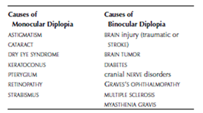
diplopia The medical term for double vision, a circumstance in which a person perceives a single object as a two distinct images. Diplopia can be vertical (images one above the other) or horizontal (images beside each other). Diplopia that is present when using both eyes and goes away when covering one EYE is binocular; diplopia that persists even when one eye is covered is monocular. Each has different clinical implications. Numerous health conditions can cause diplopia or have diplopia among their symptoms.
 The diagnostic path begins with basic OPHTHALMIC EXAMINATION and NEUROLOGIC EXAMINATION. The findings of these exams determine the direction and nature of further testing. As diplopia is a symptom rather than a condition, treatment targets the underlying cause. In degenerative disorders such as MULTIPLE SCLEROSIS and MYASTHENIA GRAVIS, diplopia may persist or worsen as the condition progresses. For monocular diplopia, patching the affected eye may alleviate the double image.
See also AMBLYOPIA; CRANIAL NERVES; STRABISMUS; VISION IMPAIRMENT.
dry eye syndrome A condition in which the lacrimal (tear) glands do not produce enough tears or the tears evaporate too quickly, causing the EYE to become dry and irritated. Dry eye syndrome has numerous causes, the most common of which are aging, medication side effects, and extended exposure to a dry or dusty environment. People who work in occupations that require close focus, such as with computers or assembly-line tasks, also may develop dry eyes as a result of insufficient blinking. Dry eyes also may accompany autoimmune conditions such as SYSTEMIC LUPUS ERYTHEMATOSUS (SLE), RHEUMATOID ARTHRITIS, and SJÖGREN’S SYNDROME. Cigarette smoking exacerbates dry eye syndrome.
The symptoms of dry eye syndrome include redness, itching, and the sensation of grit in the eyes. The diagnostic path targets identifying the underlying cause when possible. ANTIHISTAMINE MEDICATIONS, antihypertensive medications, ANTIDE- PRESSANT MEDICATIONS, and medications to treat PARKINSON’S DISEASE commonly cause dry eyes as a SIDE EFFECT; sometimes switching to a different medication reduces eye dryness and irritation.
Treatment is frequent use of artificial tears or restasis drops and remedying any identifiable cause when possible. The ophthalmologist may treat persistent dry eye syndrome with lacrimal plugs (also called punctal plugs), tiny segments of acrylic that become soft and gelatinous when inserted into the lacrimal ducts. These plugs slow the drainage of tears from the eye. Some recent studies suggest that increasing dietary intake of essential fatty acids, notably linoleic and gamma- linolenic acids, improves the eye’s ability to pro- duce tears.
See also AGING, VISION AND EYE CHANGES THAT OCCUR WITH; ALLERGIC CONJUNCTIVITIS; ALLERGIC RHINI- TIS; BLEPHARITIS; CONJUNCTIVITIS.
