THE EYES : age-related macular degeneration (ARMD) , Symptoms and Diagnostic Path , Treatment Options , Outlook and Lifestyle Modifications , Causes and Preventive Measures , aging, vision and eye changes that occur with , amblyopia , Amsler grid and astigmatism .

3396447-eye

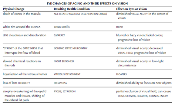
age-related macular degeneration (ARMD) A progressive condition that results in the gradual deterioration of the macula, the portion of the RETINA that provides the ability to see fine detail, and loss of vision from the center of the field of vision. ARMD is the leading cause of VISION IMPAIR- MENT, resulting in functional limitations and legal blindness in people over the age of 50. ARMD develops when the retina’s BLOOD supply diminishes. The macula’s high concentration of cones, the cells responsible for color and fine detail vision, makes it especially vulnerable to damage and its cells begin to die. The death of the cells result in diminished vision. ARMD may affect one eye at first, though nearly always affects both eyes as it progresses.

There are two forms of ARMD, atrophic (commonly known as dry) and neovascular (commonly known as wet). All ARMD begins as the atrophic form, in which the nourishing outer layer of the retina withers, or atrophies. Approximately 90 percent of ARMD remains in this form and progresses slowly. In the remaining 10 percent, new blood vessels begin to grow erratically within the choroid, the blood-rich membrane that nourishes the retina. These blood vessels are thin and fragile, and bleed easily. The resulting hemorrhages cause the retina to swell, distorting the macula and accelerating the loss of cells.

Symptoms and Diagnostic Path

ARMD begins insidiously and people tend to attribute early symptoms to the normal changes of aging. Early symptoms include

• blurring of words when reading

• “missing pieces” in the field of vision, such as parts of words or gaps in the appearance of lines or objects

• the need for increased light to perform tasks that require close vision

• faded colors

• tendency to look slightly to the side of objects to see them clearly

• distorted or wavy lines on linear objects such as signs, doorways, and railings (suggests wet ARMD)

As the macular degeneration progresses, a blind spot in the center of vision becomes apparent and enlarges. Wet ARMD progresses far more rapidly than dry ARMD. A simple screening test called the AMSLER GRID can show the gaps in vision that occur with either form of ARMD. The ophthalmologist uses further procedures, such as OPHTHAL- MOSCOPY and SLIT LAMP EXAMINATION, to visualize the retina and macula and determine which form of ARMD is present and how extensive the damage. The ophthalmologist looks for signs of exudation (swelling of the tissue that oozes fluid) that suggests wet ARMD, and for drusen (spots of depigmentation on the macula that signal the loss of retinal cells). For wet ARMD, the ophthalmologist may perform a diagnostic procedure called fluorescein angiography, in which the ophthalmologist injects fluorescein dye into a VEIN and then takes photographs of the retina as the dye flows through its blood vessels.

Treatment Options

Treatment options for ARMD are limited, and at this time there really are no treatments for dry ARMD. Some research studies demonstrate the rate of degeneration slows with increased consumption of the antioxidants lutein and zeaxanthin, and vitamins A, C, and E. For wet ARMD the laser treatments photocoagulation and photodynamic therapy are sometimes effective in sealing bleeding blood vessels and thwarting their growth, though they cannot permanently halt the neovascularization or restore vision already lost. Photo- coagulation uses a hot laser to cauterize the blood vessels but also destroys cells in the vicinity of the targeted blood vessels. With photodynamic therapy, the ophthalmologist injects a photosensitive DRUG into the person’s veins, then uses a cool laser to target blood vessels in the retina when the drug reaches them. The light of the laser is not intense enough to burn the tissue though activates the drug, which then destroys the blood vessels.

Outlook and Lifestyle Modifications

For most people who have ARMD vision declines slowly and may affect only one eye for a long time before affecting the other eye as well. Because the loss affects the center of the field of vision, vision loss is not complete though affects activities that require detailed focus, such as reading and driving, and typically reaches the level of legal blind- ness. Numerous community and health-care resources can assist with adaptive methods to accommodate diminishing vision. Even with wet ARMD, which progresses more rapidly and more severely than dry ARMD, some vision remains.

Causes and Preventive Measures Researchers do not know what causes ARMD, though it appears to have a hereditary component in that it runs in families. There are few treatments, and there is no cure, though there is evidence that antioxidants slow the rate of deterioration and the loss of vision. Vision loss is permanent. As yet there are no known measures to prevent ARMD. It appears that ARMD is more common in people who:

• smoke cigarettes

• have blue or green eyes

• experience extensive exposure to ultraviolet rays, as in sunlight exposure

• have CARDIOVASCULAR DISEASE (CVD) such as HYPERTENSION (high blood pressure), ATHEROSCLE- ROSIS, or CORONARY ARTERY DISEASE (CAD)

People who have more than one risk factor, especially when one of the risk factors is family history, should frequently and regularly monitor their vision using the Amsler grid. Early diagnosis is particularly important with wet ARMD, for which limited treatment options exist. ARMD develops in people over age 50. An ophthalmologist should evaluate changes that alter the field of vision, especially those that take the form of distortions or “missing pieces.” Regular ophthalmic examinations are important to detect ARMD as well as other conditions that affect the eye and vision with advancing age.

See also AGING, VISION AND EYE CHANGES THAT OCCUR WITH; HEMORRHAGE; OPHTHALMIC EXAMINATION; RETINAL DETACHMENT; VISION HEALTH.

aging, vision and eye changes that occur with The structures of the EYE and the processes of vision begin to undergo changes in the late fourth or early fifth decade of life. By age 65, 50 percent of people have vision impairments. By age 80, more than 90 percent of people have vision impairments. Treatment can mitigate some of these changes, such as PRESBYOPIA and CATARACT. Some conditions that affect the eye and vision develop secondary to other health conditions that are more prevalent in older people, such as DIA- BETES, HYPERTENSION, and KIDNEY disease, all of which can cause RETINOPATHY. Much loss of vision related to aging is progressive and permanent, interfering with activities such as driving, reading and other close work, and seeing at night. How- ever, most people retain the ability to see well enough to function in everyday activities.

Adaptations to accommodate the changes of the eye and vision with aging are numerous and can help maintain a desirable QUALITY OF LIFE for many people. CORRECTIVE LENSES or reading glasses are effective for presbyopia. Surgery can improve vision impairments such as cataract (CATARACT EXTRACTION AND LENS REPLACEMENT), corneal damage (corneal reshaping or CORNEAL TRANSPLANTATION), and PTOSIS and ECTROPION (BLEPHAROPLASTY). Magni-

image

fiers for reading and close work, adjustments on televisions and computers to enlarge screen images, voice-activated telephone dialers, high- intensity light sources, and screen readers with voice output are among the devices available to accommodate low vision.

See also GENERATIONAL HEALTH-CARE PERSPECTIVES; VISION HEALTH.

amblyopia A VISION IMPAIRMENT, commonly called “lazy eye,” in which the pathways between the EYE and the BRAIN do not properly handle the processes of sight. Amblyopia is most common in children. The impairment often develops when there are circumstances that allow one eye to become dominant in sending NERVE impulses to the brain, such as STRABISMUS (the inability of the eyes to focus on the same object) or congenital cataracts (opacity of the lens). Amblyopia can also develop when there is significant disparity in the refractive capabilities of the eyes, such as when one eye is hyperopic (farsighted) or myopic (near- sighted) and the other eye has normal vision. The brain becomes accustomed to messages the dominant eye and “ignores” nerve signals from the nondominant, or “lazy,” eye. Untreated amblyopia can result in permanent vision impairment or legal blindness.

The diagnostic path includes close examination of the eyes to determine whether other disease processes are present that might account for the vision deficit. Treatment targets those processes, such as cataracts or REFRACTIVE ERRORS, when they exist. When the eye is otherwise healthy and nor- mal, treatment consists of forcing the brain to rely on the amblyopic eye, usually by patching the dominant eye for structured periods of time. Sometimes the ophthalmologist will substitute atropine drops in the eye, which dilate the pupil and distort the eye’s ability to focus, when a child refuses to wear an eye patch or an eye patch is otherwise not the most appropriate therapeutic choice. The dilation interferes with the eye’s ability to focus, forcing the brain to interpret nerve messages from the untreated eye.

When detected and treated in children who are under age 9, most amblyopia responds to treatment and vision returns. Delayed or inadequate treatment may result in permanent dysfunction of the eye–brain pathways, as these become entrenched by age 9 or 10. After this time the vision pathways are well established and amblyopia can no longer develop.

See also ASTIGMATISM; HYPEROPIA; MYOPIA; PTOSIS.

Amsler grid A basic test to detect or monitor the progression of AGE-RELATED MACULAR DEGENERATION (ARMD), a condition in which the macula, the area on the RETINA responsible for fine detail vision, deteriorates. The Amsler grid is a square with evenly spaced horizontal and vertical lines, and a dot in the center of the grid. The grid’s four corners and lines should appear visible, straight, and intact. Wavy lines, gaps in the lines, or missing segments suggest damage to the macula. Such a result requires further examination from an ophthalmologist who specializes in retinal disorders.

See also VISUAL ACUITY.

astigmatism A common refractive error of vision that results from an irregularly shaped CORNEA.

Astigmatism may affect one EYE or both eyes. Typically the irregularity results in two focal points of light that reach the RETINA instead of a single focal point, resulting in blurred or distorted images. Astigmatism often coexists with HYPEROPIA (far- sightedness) or MYOPIA (nearsightedness) and tends to run in families. Corrective measures include eyeglasses, contact lenses, and REFRACTIVE SURGERY. Mild astigmatism may not produce noticeable vision disturbances, in which case it does not require correction. The success of corrective measures depends on the extent and nature of the corneal irregularities. Astigmatism often accompanies age-related changes in the eyes and vision, and is a common SIDE EFFECT of CORNEAL TRANSPLANTATION.

Less commonly astigmatism results from irregularities in the surface of LENS, called lenticular astigmatism. Options to correct for lenticular astigmatism are CORRECTIVE LENSES or lens-replacement surgery to implant an intraocular lens.

See also CATARACT EXTRACTION AND LENS REPLACE- MENT; REFRACTION TEST; REFRACTIVE ERRORS.
 

The eyes : vision impairment , health and disorders of the eyes , traditions in medical history and breakthrough research and treatment advances .

VISION IMPAIRMENT
Refractive errors occur when the focal point of light- waves entering the eye fails to align properly on the RETINA (ASTIGMATISM, nearsightedness, farsightedness).
Functional limitations result when corrected vision remains insufficient to allow participation in activities or occupations that require sight.
Legal blindness exists when corrective measures can- not restore VISUAL ACUITY to 20/200 or VISUAL FIELD to greater than 20 degrees.
Health and Disorders of the Eyes
More than 150 million Americans have a VISION IMPAIRMENT that requires CORRECTIVE LENSES (eye- glasses or contact lenses)—30 percent of men and 40 percent of women. About 12 million Americans have uncorrectable vision impairments that result in functional limitations; 10 percent of them meet the criteria for legal blindness. Among those who have uncorrectable vision impairments, 50 percent are age 65 or older. Though the eyes arise directly from the evolving brain very early in fetal development, their formation becomes complete during the final 12 weeks of PREGNANCY. Infants born before 32 weeks of gestation are at risk for RETINOPATHY of prematurity, a leading cause among children of vision impairments ranging from STRA- BISMUS (inability to focus both eyes on the same object) to legal blindness.
Traditions in Medical History
As refractive errors are very common, practitioners throughout history have tried various and sometimes hazardous methods for improving or restoring vision. The earliest documentation of corrective lenses for this purpose dates to 16th China. European traders who traveled to China noted the elderly holding quartz crystal lenses to see objects close to them. Eyeglasses set in frames and worn on the face began to appear in Europe in the 17th and 18th centuries. By the late 19th century inventors were experimenting with glass lenses placed directly on the eye. These attempts produced large, heavy, and ultimately unfeasible lenses that covered the entire surface of the eye. The contact lens finally became a reality in the 1950s with the advent of high-tech plastics that were lightweight, optically clear, and inert (did not react with body fluids). Subsequent advances over the next 30 years brought about lenses made of surgical plastics that allow oxygen to reach the cornea, much improving comfort and safety. By the 1990s, daily wear disposable contact lenses became the standard of contact lens correction.
CATARACT, the clouding and discoloration of the eye’s lens that develops with aging, has for centuries been the leading cause of blindness in adults. It also is one of the earliest documented vision problems for which practitioners used surgical treatments to remedy, perhaps because the cause of the problem, the cloudiness, was so apparent. CATARACT EXTRACTION AND LENS REPLACE- MENT has become so commonplace in contemporary ophthalmology that the procedure is no less an expectation for restoring vision than are eye- glasses for correcting refractive errors. In about 20 minutes, the ophthalmologist removes the clouded lens and replaces it with a synthetic one. Ancient physicians, lacking the benefits of the anesthetics that make the surgery painless for today’s patients, became skilled at “couching” a cataract in only seconds. The procedure required the doctor to distract the patient long enough to puncture the cornea and push the lens out of the line of vision. The lens remained within the eye as though resting, hence the term “couching.” The result was less than perfect because the person lost the refractive ability of the lens, but the procedure restored enough vision to allow one to function in daily life. In the 1950s ophthalmologists began removing the cataract from the eye, but not until the 1970s did technology and technique converge in procedures that incorporated a replacement lens.
Breakthrough Research and Treatment Advances The evolution of knowledge and advances in laser technology are converging to present treatment options that were science fiction a decade ago. New procedures are greatly expanding the potential for permanent correction of disorders and defects of the eye, including refractive disorders, that reduces and may eventually even eliminate the need for corrective lenses. Refined laser techniques such as LASIK allow ophthalmologists to reshape the cornea in precise, microscopic increments. Implantable rings inserted around the edge of the cornea can help flatten and reshape it to alter its refractive ability. Permanent contact lenses attached over the lens can have similar effect. Implantable replacement lenses are expanding beyond their initial application in cataract extraction and replacement to offer nearly ideal vision for people with severe astigmatism or myopia (nearsightedness).
Cataract extraction and lens replacement now routinely restores sight for more than 90 percent of people who otherwise would lose vision to cataracts. Other surgical procedures offer hope for altering the course of glaucoma. New treatments may stem the loss of vision due to AGE-RELATED MACULAR DEGENERATION (ARMD). These conditions are the leading causes of vision impairments that lead to functional limitations or legal blindness among adults. And research continues to explore a “bionic” PROSTHETIC EYE that can convert light- waves to nerve impulses and transmit them to the brain. Such a prosthesis would function similarly to the COCHLEAR IMPLANT used to restore some types of neurosensory HEARING LOSS. Because many of the conditions that result in vision impairment are not preventable, technological innovations such as these appear to be the future of ophthalmologic treatment.
 

THE EYES : Structures of the Eye , Functions of the Eye , MUSCLES THAT MOVE THE EYE and The physics of vision .

THE EYES

The eyes conduct the function of vision. Practitioners who provide care for the eyes and vision may be ophthalmologists (medical doctors who specialize in ophthalmology, providing medical and surgical treatment for diseases of the EYE) or optometrists (doctors of optometry who specialize in diagnosing and correcting REFRACTIVE ERRORS of vision). This section, “The Eyes,” presents a discussion of the structures of the eye and how they function to provide the sense of sight, an overview of VISION HEALTH and disorders, and entries about the health conditions that can affect the eyes and vision.

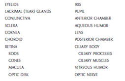
Structures of the Eye

image

Functions of the Eye

Ancient philosophers viewed the eyes as the windows to the soul, based on the belief that the PINEAL GLAND, located deep within the BRAIN, held the soul. Their rudimentary understanding of anatomy and physiology led them to conclude that the optic nerves connected the pineal gland and the soul directly to the outside world through the eyes. Though modern knowledge of the body’s structure and function clarifies that no such physical pathway exists, ancient scientists were not entirely off track. The pineal gland does appear to receive direct information from the external environment, which influences its production of MELATONIN, a HORMONE related to the body’s circadian cycles (cycles of wakefulness and sleep). Researchers do not fully understand the mechanisms of this, and it is possible the OPTIC NERVE plays some role. However, the primary function of the optic NERVE is to provide a direct conduit from the EYE to the brain through which the brain receives about two thirds of the information it processes about the environment out- side the body.

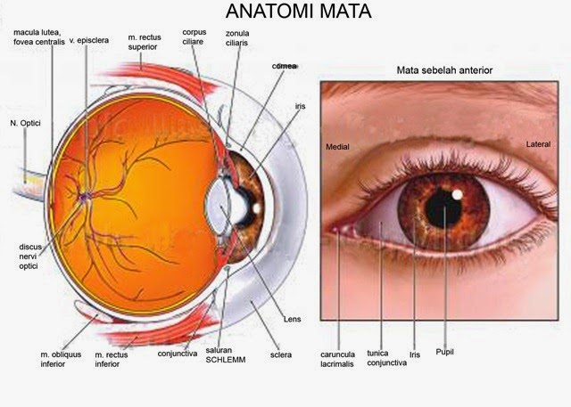
The eye resides within the protective enclosure of the orbit, a socket of BONE in the skull. Thin pads of fat cover the orbital bones to cushion the eye. A small opening in the back of the orbit allows passage of the optic nerve and the blood vessels that supply the eye. The eyelids, upper and lower, blink—automatically open and close—15 to 20 times a minute to rinse the eye with tears. Reduced blink rate is a characteristic of neurologic disorders such as PARKINSON’S DISEASE; increased blink rate occurs with eye irritation such as CON- JUNCTIVITIS and diseases such as MENINGITIS. The tears then drain from the lacrimal sac at the inner corner of the eye into the upper NOSE. The eyelids also close to protect the eye from hazards such as foreign objects and very bright light, and to cover the eye during sleep to keep it moist. The eye- lashes, extending from the eyelids, also help keep foreign objects from striking the eye and the eye- brows channel sweat around the eyes.

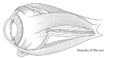
Six muscles attach the eye to the orbit, functioning in pairs as well as in coordination with one another to move the eye. These muscles integrate into the sclera, the fibrous outer layer of the eye, and extend to the back of the orbit where they anchor to the bone. When one MUSCLE in a pair contracts, the other relaxes. Typically both eyes move in tandem, which allows the eyes to simultaneously focus on the same object. This

image

imageprovides depth perception and accommodates each eye’s “blind spot.” Some people have the ability to intentionally move their eyes independent of each other, though unintentional disparate movement generally indicates a pathologic condition. Discordant movement may characterize neurologic disorders such as progressive supranuclear palsy (PSP) and TRAUMATIC BRAIN INJURY (TBI).

Abnormal eye movements also accompany vestibular disorders (disturbances of the balance mechanisms of the inner EAR).

MUSCLES THAT MOVE THE EYE

Superior oblique and inferior oblique rotate the eye primarily in a circular motion.

Superior rectus and inferior rectus move the eye primarily up and down.

Lateral rectus and medial rectus move the eye primarily side to side.

How the eye “sees” The sclera gives the eye its shape and rigidity. The front part of the sclera forms the “white” of the eye, the coloration coming from the white pigmentation of the fiber cells. In its center, the sclera becomes transparent, forming the CORNEA. The middle layer of the eye’s wall is the choroid, a thin, dark membrane rich in BLOOD vessels. The choroid loosely attaches to and nourishes the sclera and the eye’s innermost layer, the RETINA, where sight becomes vision.

Specialized cells infuse the retina, which lines the back of the inner eye. These cells, rods and cones, convert lightwaves into electrical impulses. Rods are the most plentiful, numbering about 120 million on each retina, and detect light in perceptions of shades of gray. Cones detect color and detail; there are about 6 million of them on each retina. Cones are sensitive to red, green, or blue. Rods and cones contain photosensitive chemicals that react to different wavelengths of light. The reactions alter the electrical charges of the rods and cones, creating nerve signals. Each minute of wakefulness thousands of these impulses traverse the optic nerves, carrying messages the brain then interprets and assembles as visual images.

The optic nerve, which contains more than a million nerve fibers, carries these signals to the brain. The pigmented cells of the retina are rich in melanin, the same chemical that causes the SKIN to darken in response to sun exposure. In the retina, these cells form a “blackout screen” that eliminates reflection, allowing lightwaves to reach and activate the rods and cones without interference. The macula, a small circular area in the center of the retina, contains the most dense distribution of cones and handles fine detail vision. The “blind spot,” the point at which the optic nerve enters the retina, is the optic disk; it contains no rods or cones. RETINITIS PIGMENTOSA (hereditary degeneration of the retina) and RETINAL DETACHMENT (separation of the retina from the choroid) are among the conditions that can affect the retina, resulting in impaired vision and blindness.

The physics of vision Lightwaves pass through the cornea and the LENS to enter the eye through the pupil, the opening in the circular muscle that rings the lens, the iris. The iris is the colored part of the eye; the pupil in its center appears black because it reveals the dark interior of the eye. The iris dilates (increases the size of) the pupil to allow more light to enter the eye and constricts (decreases the size of) the pupil to reduce the light that enters the eye. The cornea and the lens each refract, or bend, the entering lightwaves. The ciliary muscles contract and relax to move the lens, which thickens or flattens, respectively, to improve focus. After about age 40 the lens gradually loses its FLEXIBILITY, accounting for age-related difficulty with near vision (PRESBYOPIA).

Refracted light forms a final focal point that, in the healthy eye, aligns in a pattern on the retina at the back of the eye. The mechanics of this refractory process are such that the image result- ing on the retina is upside down. When interpreting and assembling nerve signals from the eye, the brain automatically reverses the image to perceive it right-side up. Refractive ASTIGMATISM, HYPEROPIA, and MYOPIA when the final focal point falls short of or extends beyond the retina, resulting in images that are out of focus or distorted.

Helping keep the lightwaves from fragmenting during refraction are two chambers of fluid, the aqueous humor, which fills the space between the cornea and the lens (the anterior chamber), and the vitreous humor, which fills the interior of the eye. The ciliary processes, specialized folds of the eye’s choroid layer that extend into the posterior chamber at the corners of the lens behind the iris, produce aqueous humor. This watery fluid is about the consistency of saliva and serves also to lubricate and nourish the cornea. Aqueous humor circulates through the anterior chamber between the cornea and the lens, then drains from the eye via the drainage angle, a channel between the iris and the cornea. Dysfunction of the drainage angle is a hallmark characteristic of GLAUCOMA.

Vitreous humor forms when the eye completes its development during the final trimester of gestation. A substance similar to water in chemical composition and to gelatin in consistency, vitreous humor maintains the eye’s shape and helps keep the retina smooth and even against the back of the eye. The volume of vitreous humor increases as the eye grows though otherwise remains constant (unlike the aqueous humor, which the eye continuously produces). Around age 40 years the vitreous humor begins to liquefy as a normal process of aging, causing VITREOUS DETACHMENT, which usually has little effect on vision though can produce FLOATERS (fragments of tissue that become suspended in the vitreous humor).


Ten Ways to Go From Day to Night in a Flash : Leaving the house in the morning ready for work and play , Transformers aren’t just toys and Turning day into night with accessories

Ten Ways to Go From Day to Night in a Flash

In This Chapter

▶ Leaving the house in the morning ready for work and play

▶ Transformers aren’t just toys

▶ Turning day into night with accessories

Whether you’re heading to a planned event or a last-minute soiree, you want to look your best, even when you don’t have the time you’d like to primp and polish. No worries. In this chapter, I give you ten ways to trans- form your look from day to night. With these tips, you’ll look like you spent hours digging through your closet to find the perfect outfit!

From Simple to Sizzling

If you know you’re going out after work, slip on a simple black dress in the morning with whatever shoes are office-appropriate. Take along some jewelry, a clutch, and a pair of dressy sandals. When your work day is done, all you need to do to totally transform your look is put on the jewelry (the right amount of bling can spice up that little black dress), trade in your day bag for the clutch, and don your dressy sandals (see Figure 18-1). In no time flat, you have a whole new look!

clip_image007

Figure 18-1: The little black dress takes you from day to night in a flash.

A Silky Transformation

Simply by changing the way you wear a silk scarf, you can change your whole look. All it takes is a little creativity, a few seconds, and the right scarf (a 36-inch square works, or if you want more coverage, go bigger). During the day, tie the scarf around your neck and pair it with a suit and tank. At night, take the scarf off your neck, remove the tank and suit jacket, and tie the scarf as a halter top (see Figure 18-2). Chapter 15 explains how to turn a scarf into a halter. Who knew a simple scarf could take you so far?

clip_image009

Figure 18-2: A little creativity with a scarf gives you a new look almost instantly.

Cubicle to Cocktails

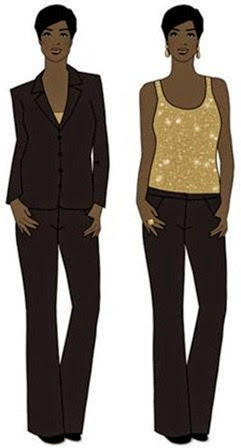
If you’re going out after work, there’s no need to pack an extra outfit. Simply wear a suit. Underneath, put on a sexy tank that barely shows when the jacket is closed (see Figure 18-3). When you’re ready to go meet your friends for cocktails, take off your jacket and you’ll be ready for that cosmo!

clip_image011

Figure 18-3: Less can be more when dressing up for an evening out.

A Shirt to Take You Anywhere

A white button-down shirt is suitable for almost any occasion. During the day, pair it with jeans. At night, put it with a dressier skirt or slacks. Add a bold necklace or chandelier earrings for a little pop (see Figure 18-4). The look is not only sophisticated, but understated and sexy as well!

clip_image013

Figure 18-4: Trade in your slacks for a dressy skirt when going out on the town!

Tote to Clutch

If you’re heading to a party after work, don’t let your bag be the sign that you didn’t have time to run home and change. An overflowing tote is never stylish. To avoid this dilemma, throw a clutch in your purse (pack some mini- makeup in your evening bag, too). That way when you’re running out of the office for your fabulous plans, you’ll look chic from head to toe!

Accessorize, Accessorize!

Never underestimate the power of accessories. If you’re in a simple dress during the day and want to dress it up for dinner, you don’t need to change your outfit;

you can simply change your accessories. Pick two pieces to make your outfit pop (shoes don’t count!). Choose a bracelet and earrings, a necklace and statement ring, or a pair of earrings and a belt (as pictured in Figure 18-5). Any such combo will do the trick. Have fun with your choices, and don’t be afraid to mix metals. The only caveat? Don’t overdo it. (See Chapter 15 for details on how to avoid over-accessorizing.)

clip_image015

Figure 18-5: The right accessories can dress up any outfit.

Shoulder Bag to Clutch

Invest in a bag that has a chain or strap you can take off or tuck inside. If you’re shopping during the day and want to be “hands-free,” you can use the strap. At night, when you just want to wear a clutch, you can tuck the strap inside the bag and voila! Most major department stores carry these types of bags. With most chain-strap bags you can tuck in the strap. Look around and find one you can wear with many different outfits; it’s a great staple for any wardrobe.

Day Casual to Dinner Chic

A simple sweater set is a great staple. Paired with a pair of slacks, it’s always classic and clean — a great look for the office. Dress it up for night by taking the cardigan off, tying it around your neck, and pairing it with a fun skirt, sandals, a clutch, and accessories (see Figure 18-6). Now you’re ready to dance the night away in style!

clip_image017

Figure 18-6: Maybe you never thought you could use a sweater as an accessory, but you can.

It’s a Wrap!

You can wrap many dresses in different ways to get different looks. American Apparel makes a great and inexpensive one that’s very easy to figure out and experiment with. Don’t be afraid to come up with your own way to wrap a versatile dress…whatever works best on your body works! Here’s an example: For daytime, wrap the two straps of the dress around your waist and tie a knot in the back (I wear this look all the time). Pair with a T-shirt and cardigan and you’re ready to shop the day away! For nighttime, tie the two straps around your neck to make a halter dress (see Figure 18-7). Get out those

fab new earrings you’ve wanted to wear and a pair of high heels, and you’re ready to dance into the night!

Best Foot Forward

Higher heels are sexy and great for nighttime, but few women can walk around in them comfortably all day. You could bring another pair of shoes with you, but there’s another solution: Camileon Heels, shoes that, with a simple move, have a heel that goes from low to high in a flash (see Figure 18-8). And they look nice, too! To buy a pair, go to www.camileonheels.com.

image

©Richard Quindry, Camileon Heels

Figure 18-8: With these shoes, you switch from low to high heels in no time.


Ten Fashion Faux Pas and How to Avoid Them : Knowing what to show and what not to show and Avoiding other mistakes

Ten Fashion Faux Pas and How to Avoid Them

In This Chapter

▶ Knowing what to show and what not to show

▶ Avoiding other mistakes

Faux pas is a French expression that in English means something you’re not supposed to do. In this chapter, I list ten fashion mistakes you want to avoid at all costs.

Over-Accessorizing

You’ve heard the old saying “less is more,” right? Like many old sayings, it’s lasted for a reason. Wearing big statement pieces to express your personal style is definitely fun and fashionable, but you don’t want to wear too many statement pieces at one time. Pick one piece that you want people to notice. Wear a chunky necklace, chandelier earrings, or a cute hair accessory. Wearing all of them at once overwhelms your overall look, distracts anyone from looking at your face, and makes you look like you tried too hard to be matchymatchy. (For more information on how to accessorize, see Chapter 15.)

Problematic Panties

Doesn’t the title say it all? Panties should always be worn under, not out of, your clothing. Obviously, panties sticking out of your jeans or slacks is a giant no-no. Make sure (and you can even test this) that no matter what your pose — sitting, standing, or bending to pick up a pen — your undergarments don’t expose themselves (see Figure 17-1).

Your jeans or pants are there to cover your rear and flatter it. If your under- wear is sticking out, chances are good that your outfit is doing nothing for your entire midsection and backside. Don’t draw unwanted attention there by letting your panties, or worse, your rear, stick out of them. Yikes!

Wearing White to a Wedding

As is tradition, the bride will (most likely) be in white, so you shouldn’t be. In addition to white, avoid ivory, cream, ecru, eggshell, linen, or any other nearly white color. People often question whether black is okay to wear to a wedding. If it’s a nighttime affair, then yes, black is totally chic. However, if it’s a daytime affair, stay away from black. For more on dressing for special occasions, head to Chapter 11.

Baring Your Stomach

Baring your stomach — unless you’re wearing a bathing suit — is another giant no (see Figure 17-2). Just as showing your rear isn’t the height of fashion sophistication, neither is showing your stomach. Even if you can get beyond the lack of sophistication, it’s not a flattering look for most women. A cropped top cuts you right in the middle and draws all the attention to your waist. Believe me, you can look sexy in many ways; showing your stomach is not one of them.

Wearing Hose with Open-Toe Shoes

Gasp! Seeing the seam of pantyhose across someone’s toes in an open-toe shoe gives me hives! If you’re wearing sandals, it’s probably warm out, and you don’t need hose. If you’re wearing pantyhose, it’s probably cold enough out to warrant closed-toe shoes. Even if you came up with what seemed like a logical reason to pair hose and open-toe shoes, the look is an absolute no. No exceptions! Head to Chapter 14 for more hosiery rules.

Part of being fashionable is dressing appropriately for the season and the weather. If it’s hot, wear sexy sandals and go with bare legs. If it’s cold, pair the hose or tights with great boots or pumps. Of course, you can also wear open-toe sandals (sans hose) in the winter with an evening dress, as long as you’re not outdoors for an extended period.

Wearing High Waters

Wearing capri pants is one thing; wearing pants that are meant to be full length but that fall too high on your leg is quite another (see Figure 17-3). Make sure your pants hit the top of your shoe (or foot, if you’re wearing a sandal). You don’t want to look like you’re ready for a flood.

Wearing pants that are too short not only takes away from the long, clean line that is supposed to make you appear taller and thinner, but it also looks like you bought the wrong size, got them hemmed improperly, or shrank them in the wash — none of which is good.

Too much

The most common fashion faux pas is to overdo it. That could mean wearing too much jewelry, too much perfume, too much makeup; show- ing too much skin; or wearing too tight a dress or too high a heel. You may think that, because so many of the outfits seen on the fashion runways are over the top, exaggeration has a part to play in everyday fashion, but that’s not so. Remember, all fashion is an accessory to the person wearing the clothes, you. If you overdo it, then you will get overwhelmed. So while you always want to make a style statement, you never want to be a fashion victim. If you abide by the golden rule — less is more — you’ll always be able to avoid becoming a fashion faux pas.

clip_image012

Figure 17-3: Pants aren’t supposed to look like they shrank in the wash.

Wearing White Underwear under Anything Sheer

If you’re wearing a sheer top, choose flesh-toned undergarments. These come in a variety of shades, so you can find one that matches your skin tone. The flesh-toned color of your bra, camisole, or slip gives the illusion that you don’t have anything on underneath, while everything really remains covered and hidden. This way, you get the full effect of the beauty of the sheer top or dress without the distraction of the wrong undergarment. When you wear white underneath something sheer, the white is all you see (see Figure 17-4). This is especially true if you’re somewhere where you may be photographed — such as a wedding. The flash of the camera will pick up the white undergarment, making it even more noticeable in the picture than it is in real life.

Wearing All Denim

I love denim. Jeans are a great staple in most everyone’s wardrobe. A jean jacket is the perfect complement to white pants, khakis, or a cute dress.

Still, don’t wear jeans and a denim jacket together as an outfit. The look is straight out of the 1980s and is too matchy-matchy. With so many mix and match options for jeans and jean jackets, pair denim with pretty much anything other than more denim. (For more on how to wear denim, see Chapter 5.)

Showing Your Bra Straps

Your bra straps should never be showing. If you’re wearing a spaghetti strap dress, opt for a strapless bra. If you’re wearing a racerback tank top, go for a racerback bra. Letting your straps show takes away from your outfit, and your bra straps are often the first thing people notice (see Figure 17-5). You can find convertible bras in all different sizes and with all different levels of support. (Head to Chapter 14 for more on undergarments.)

Many bra companies have begun to make fashionable bra straps that are meant to be shown. This look is very casual and therefore inappropriate for work and many other occasions. Exercise caution with fashionable straps, and remember, a cleaner look is always safe and stylish.

Wearing Clothing That’s Too Tight

Clothing is unflattering when it’s too tight. I know, I know — you love that top and you must have it. Unfortunately, the only size you can find is a size smaller than you wear. What do you do? You put the garment down and walk away! We’ve all bought the top we had to have knowing it was too small. But no matter how fab the top, if you have to squeeze yourself into it, it’s a total no. Walking around in clothing that’s too tight is not only uncomfortable; it’s also unfashionable (see Figure 17-6). You don’t want to be the woman in a fabulous outfit that’s too small.