The eyes : keratitis and keratoconus

images (4)

keratitis INFLAMMATION of the CORNEA, usually the result of an INFECTION. The cause of the infection is more commonly viral, such as HERPES SIMPLEX or HERPES ZOSTER, than bacterial. Symptoms of infectious keratitis include

• redness and irritation of the EYE and conjunctiva (inner eyelids)

• discomfort or PAIN

• excessive tearing

• difficulty keeping the eye open

• diminished VISUAL ACUITY (usually blurred vision)

• eye discharge or crusting

Viral keratitis usually runs its course without complication, though occasionally a secondary bacterial infection may develop. ANTIVIRAL MEDICA- TIONS sometimes shorten the course of chronic herpes infections. Bacterial keratitis typically follows a CORNEAL INJURY, such as an abrasion or laceration, and requires treatment with ophthalmic ANTIBIOTIC MEDICATIONS. Chronic or recurrent keratitis can cause permanent scarring of the cornea, resulting in diminished visual acuity such as ASTIG- MATISM. Extensive corneal damage may require CORNEAL TRANSPLANTATION.

SUNBURN is the most common cause of noninfectious keratitis. Extended exposure to the sun, especially on or around water, exposes the surface of the eye to the same ultraviolet rays that cause sun- burn of the SKIN. Ultraviolet burns to the cornea are painful; treatment with ophthalmic CORTICOSTEROID MEDICATIONS helps reduce the inflammation.

See also BACTERIA; CONJUNCTIVITIS; EPISCLERITIS; IRITIS; SCLERITIS; SUN PROTECTION; UVEITIS; VIRUS.

keratoconus A degenerative disorder in which the CORNEA thins, allowing it to protrude from the surface of the EYE in somewhat of a cone shape. Keratoconus affects both eyes though often progresses at different rates in each eye. Ophthalmologists do not know what causes keratoconus, though it appears to run in families. Keratoconus is painless though results in progressive VISION IMPAIRMENT, typically in the forms of MYOPIA (near- sightedness) and ASTIGMATISM (distortions of vision resulting from the irregular surface of the cornea). As these REFRACTIVE ERRORS are the primary symptoms, the keratoconus may not become apparent until the coning becomes obvious.

Treatment for mild to moderate keratoconus is rigid gas-permeable contact lenses, which correct the refractive errors of vision as well as help contain the shape of the cornea. As the keratoconus progresses, however, contact lenses become less effective, and the thinned cornea may not be able to tolerate them. Keratoconus ultimately destroys the cornea and is a leading reason for CORNEAL TRANSPLANTATION, which replaces the diseased cornea with a donor cornea. Corneal transplantation successfully restores vision in about 90 per- cent of people who have keratoconus and undergo the procedure.

In 2004 the US Food and Drug Administration (FDA) approved a new treatment for keratoconus, corneal inserts, which are tiny plastic rings the ophthalmologist implants along the edge of the cornea. The corneal inserts flatten the cornea, reducing the coning. Corneal inserts come in different thicknesses to allow less or more flattening and are replaceable.

See also CORNEAL INJURY; CORRECTIVE LENSES


The eyes : lens , mydriasis and myopia .

173237-1

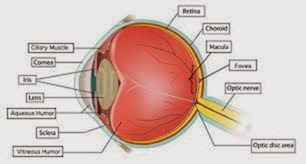
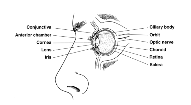
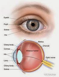
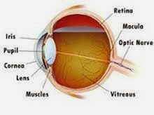
lens The primary focusing structure of the EYE, located in the center at the front of the eye. The lens is transparent, convex (rounded outward on each side), round, and flexible. A thin membrane encloses the lens. Tiny muscles at the front edges of its sides, the ciliary muscles, contract to flatten the lens and relax to thicken the lens. These adjustments alter light refraction (the angle at which the lens bends lightwaves entering the eye) to accommodate near and distant vision. The most common health conditions that affect the lens are PRESBYOPIA, in which the FLEXIBILITY of the lens diminishes with aging, and CATARACT, in which protein deposits cloud the lens and obscure vision. The lens is also vulnerable to accidental injury, particularly from blunt force (such as a baseball) or puncture.

For further discussion of the lens within the context of ophthalmologic structure and function please see the overview section “The Eyes.”

See also CATARACT EXTRACTION AND LENS REPLACE- MENT; CORNEA; HYPEROPIA; MYOPIA; REFRACTIVE ERRORS; RETINA; VISION IMPAIRMENT.

mydriasis Excessive or persistent dilation of the pupil that is a symptom of ophthalmic or systemic conditions. The ophthalmologist may induce mydriasis, using topical atropine, to examine the inner EYE. Therapeutic mydriasis using atropine- based ophthalmic drops is an alternate treatment for AMBLYOPIA. Two eye conditions can cause mydriasis: GLAUCOMA and damage to the iris. In the healthy eye the iris, a muscular membrane, con- trols the opening of the pupil. INFLAMMATION of or tears in the iris affect its ability to function, which also can result in mydriasis.

Other causes of mydriasis are systemic, involving damage to NERVOUS SYSTEM structures and functions, and may include TRAUMATIC BRAIN INJURY (TBI), STROKE, and medication response such as with narcotic use, which causes the muscles to relax. Eye disorders often affect only one eye (unilateral mydriasis), whereas systemic conditions typically affect both eyes (bilateral mydriasis). PHOTOPHOBIA (sensitivity to bright light) often accompanies mydriasis as the dilated pupil cannot limit light from entering the eye. VISION IMPAIRMENT depends on the extend of the dilation; a fully dilated pupil prevents focus on near objects.

The diagnostic path begins with a basic OPH- THALMIC EXAMINATION including SLIT LAMP EXAMINATION and OPHTHALMOSCOPY, unless there is clear evidence that the mydriasis results from systemic causes. TONOMETRY, which measures the pressure within the eye (INTRAOCULAR PRESSURE), determines whether glaucoma is present. Tears of the iris are typically apparent when looking at the eye as they distort the iris (the colored portion of the eye). Inflammation of the iris (IRITIS) often reddens the eye and is apparent with ophthalmoscopy. Further diagnostic measures turn to NEUROLOGIC EXAMINA- TION. Treatment targets the causative condition.

See also EYE PAIN; NARCOTICS; TRAUMA TO THE EYE.

myopia A refractive error commonly called nearsightedness, in which the EYE has difficulty focusing on distant objects. Myopia results when the focal point of lightwaves entering the eye falls short of the RETINA, causing the images the retina registers to be blurred. The refractive error occurs because the distance from the front to the back of the eye is longer than normal. Symptoms of myopia include

• squinting when looking at distant objects

• straining to see when driving

• difficulty seeing the ball when playing baseball, tennis, and similar sports

• frequent headaches at the end of the day

CORRECTIVE LENSES (eyeglasses or contact lenses) can compensate for myopia by altering the focal point of lightwaves so it falls directly on the retina. They do so by refracting, or bending, the light- waves inward. Eye professionals denote refractive corrections in units of measure called diopters. For myopia, the expression of diopter is a negative number. REFRACTIVE SURGERY, which permanently

alters the shape of the CORNEA, provides refractive correction for mild to moderate myopia (–1 to –15 diopters). In 2004 the US Food and Drug Administration (FDA) approved an implantable contact lens to improve severe myopia (–15 to –30 diopters). Severe myopia sometimes cannot be fully corrected, resulting in VISION IMPAIRMENT with functional limitations or legal blindness. Myopia is the most common refractive error, affecting about 35 percent of adults.

See also ASTIGMATISM; HYPEROPIA; PRESBYOPIA; REFRACTIVE ERRORS.


The eyes : intraocular pressure , iritis and ischemic optic neuropathy .

images (3)

intraocular pressure The pressure within the EYE that maintains the eye’s form and structure. Nor- mal intraocular pressure in an adult is 12 to 22 millimeters of mercury (mm Hg). A device called a tonometer measures intraocular pressure, either through light contact against the anesthetized eye or via the force of resistance to a puff of air blown against the eye’s surface (a noncontact method that does not require anesthetic drops). Elevated intraocular pressure is called ocular hypertension. Lower than normal intraocular pressure is called ocular hypotension.

Ocular hypertension (greater than 21 mm Hg) is a hallmark sign of GLAUCOMA, an eye condition that, if untreated, results in complete loss of vision. Other health conditions that can increase intraocular pressure include tumors that press against the eye, ORBITAL CELLULITIS, and GRAVES’S OPHTHALMOPATHY. Increased intraocular pressure damages the cells on the front of the OPTIC NERVE (the retinal ganglia), leading to permanent VISION IMPAIRMENT. Ophthalmic medications that reduce intraocular pressure work through various mechanisms, depending on the cause of the increased pressure.

Ocular hypotension, in which the intraocular pressure is lower than normal (less than 12 mm Hg), characterizes chronic UVEITIS (INFLAMMATION of the structures of the eye) and of certain tumors of the eye. Ocular hypotension also sometimes accompanies systemic HYPOTENSION (low BLOOD PRESSURE) and as a SIDE EFFECT of medications, notably general anesthesia agents.

See also OPHTHALMIC EXAMINATION; TONOMETRY; VITRECTOMY; VITREOUS DETACHMENT.

iritis INFLAMMATION of the iris, the MUSCLE surrounding the pupil of the EYE. Iritis may develop

as a result of INFECTION, such as CONJUNCTIVITIS that spreads to involve other structures of the eye. Iritis also occurs as part of the inflammatory process in AUTOIMMUNE DISORDERS such as RHEUMATOID

ARTHRITIS. The symptoms of iritis include

• irritation and a “gritty” sensation in the eye

• redness of the eye

• PHOTOPHOBIA (sensitivity to bright light)

• blurry vision

• excessive tearing

The ophthalmologist can diagnose iritis based on the appearance of the iris and the eye, though will additionally perform a SLIT LAMP EXAMINATION to look for involvement of other structures of the eye. Treatment is ophthalmic ANTIBIOTIC MEDICA- TIONS (eye drops or ointment) when the ophthalmologist suspects bacterial infection and ophthalmic CORTICOSTEROID MEDICATIONS to reduce the inflammation. When the cause of the inflammation is systemic, the ophthalmologist may pre- scribe anti-inflammatory medications such as corticosteroids or NONSTEROIDAL ANTI-INFLAMMATORY DRUGS (NSAIDS). Treatment usually resolves the symptoms without residual VISION IMPAIRMENT. Untreated or recurrent iritis can have long-lasting effects on vision, including increased INTRAOCULAR PRESSURE.

See also BACTERIA; EPISCLERITIS; GLAUCOMA; KER- ATITIS; SCLERITIS; UVEITIS.

ischemic optic neuropathy Damage to the OPTIC NERVE resulting from insufficient blood supply, sometimes called “STROKE” of the optic nerve. Ischemic optic neuropathy is occurs most commonly in people over age 55 and causes mild to complete VISION IMPAIRMENT. There are two forms: arteritic, associated with giant-cell arteritis (an inflammatory disorder of the arteries that typically affects the temporal arteries) and nonarteritic, which correlates with CARDIOVASCULAR DISEASE (CVD) such as HYPERTENSION (high BLOOD PRESSURE) and ATHEROSCLEROSIS. Other conditions associated with the nonarteritic form include DIABETES, HYPOTENSION (low blood pressure), and RHEUMATOID ARTHRITIS.

Vision impairment due to ischemic optic neuropathy comes on suddenly. In a characteristic pat- tern, a person wakes up in the morning with noticeable loss of VISUAL ACUITY and VISUAL FIELD. This may continue for several days, improving as the day goes on, though in short order becomes permanent. The diagnostic path begins with OPH- THALMOSCOPY and SLIT LAMP EXAMINATION to visualize the optic disk (portion of the optic nerve that attaches to the RETINA), which appears pale and swollen. Diagnosis of giant cell arteritis by temporal ARTERY biopsy confirms the diagnosis of the arteritic form of ischemic optic neuropathy. Doctors arrive at the diagnosis of the nonarteritic form on the basis of symptoms and ruling out other causes.

Treatment for arteritic ischemic optic neuropathy is CORTICOSTEROID MEDICATIONS to reduce the INFLAMMATION. Vision loss, however, is irreversible. There are no effective treatments for the nonarteritic form, which does not appear to improve with corticosteroids. Lifestyle modifications such as SMOKING CESSATION improve circulation in general with the presumption that such improvement also affects optic structures. ASPIRIN THERAPY, such as prescribed as a prophylactic measure for HEART ATTACK and stroke, may have a preventive effect with the arteritic form. It is especially important to manage underlying health conditions that affect circulation, such as hypertension and diabetes.

When the ischemic optic neuropathy affects only one EYE, the person can make adaptations and adjustments to accommodate the vision impairment that do not necessarily require substantial changes in lifestyle. Most people can still read, work, and perform other functions of daily living with visual acuity in only one eye. Ischemic optic neuropathy that involves both eyes can significantly affect lifestyle.

See also TOXIC OPTIC NEUROPATHY; VASCULITIS.


The eyes : hordeolum and hyperopia .

images (2)

hordeolum A bacterial INFECTION of a gland or an eyelash follicle along the edge of the eyelid, commonly known as a stye. A hordeolum causes swelling, redness, PAIN, and a discharge that leaves a crusty layer on the eyelids during sleep. The doctor can usually diagnose hordeolum by its presenta- tion. BLEPHARITIS (infection of the inside surface of the eyelid) and CONJUNCTIVITIS (infection of the conjunctiva, the membrane lining the eyelids) may instigate, accompany, or follow hordeolum. The doctor often chooses to anesthetize the area and lance (make tiny punctures or incisions under sterile conditions) the hordeolum to drain its contents and relieve the pressure. Further treatment is ophthalmic ANTIBIOTIC MEDICATIONS, typically in ointment form, applied to the area. Sometimes oral antibiotic medications are also necessary. Warm, moist compresses soothe the irritated tissues and help draw out any remaining pus.

Most hordeola clear up in 7 to 10 days with treatment and heal without residual consequences. A hordeolum does not itself cause VISION IMPAIRMENT, though untreated hordeola can lead to significant EYE problems if the infection spreads to other structures of the eye. Some people experience recurring hordeola, while others experience only a single episode. A hordeolum may also form the basis for a CHALAZION (painless nodule) to develop in its place.

See also BACTERIA; ECTROPION; ENTROPION.

hyperopia A refractive error, commonly called farsightedness, in which the EYE has difficulty focusing on near objects. Hyperopia results when the focal point of lightwaves entering the eye extends past the RETINA, causing the images the retina registers to be blurred. The refractive error occurs because the distance from the front to the back of the eye is shorter than normal. Symptoms of hyperopia include

• squinting when reading or doing close work

• HEADACHE

• fatigued eye muscles (aching around the eyes)

• blurred vision when looking at near objects yet clear vision when looking at distant objects

CORRECTIVE LENSES (eyeglasses or contact lenses) can compensate for hyperopia by altering the focal point of lightwaves so it falls directly on the retina. They do so by refracting (bending) the lightwaves outward. REFRACTIVE SURGERY, which permanently alters the shape of the CORNEA, can provide refractive correction for people with mild to moderate hyperopia. Hyperopia sometimes occurs following refractive surgery for MYOPIA (nearsightedness) as a consequence of overcorrection. Eye professionals denote refractive corrections in units of measure called diopters. For hyperopia, the expression of diopter is a positive number. Corrective lenses for hyperopia have a magnifying appearance that make the eyes look bigger than they are.

Hyperopia is less common than myopia, affecting about 20 to 25 percent of adults. Few people who have hyperopia have greater than +6 diopters of refractive error, so nearly always corrective measures result in normal VISUAL ACUITY.

See also ASTIGMATISM; PRESBYOPIA; REFRACTIVE ERRORS.


The eyes : glaucoma , Symptoms and Diagnostic Path , Treatment Options and Outlook , Risk Factors and Preventive Measures , Graves’s ophthalmopathy , Symptoms and Diagnostic Path , Treatment Options and Outlook and Risk Factors and Preventive Measures

glaucoma A serious and progressive EYE condition in which the cells at the front of the OPTIC NERVE where it intersects with the RETINA, the retinal ganglia, die, resulting in vision loss. Early diagnosis and treatment can minimize vision loss. Health experts estimate that 5 million Americans have glaucoma, though only about 2 million of them know it. Glaucoma is the third-leading cause of blindness in the United States, primarily because it remains undetected until damage to the optic NERVE becomes significant. Glaucoma becomes more common after age 65, though there is a congenital form that manifests in early child- hood (congenital glaucoma).

Until the late 1990s ophthalmologists perceived glaucoma to be the exclusive consequence of increased pressure within the eye (INTRAOCULAR PRESSURE) that caused the death of retinal ganglia cells. Current understanding of the disease process of glaucoma affirms the death of retinal ganglia cells as the cause of damage to the optic nerve, though recognizes that numerous factors, intraocular pressure being only one among them, con- tribute to this damage. About 30 percent of people who have glaucoma have normal intraocular pressure, and only about 10 percent of people who have elevated intraocular pressure have glaucoma.

There are two general forms of glaucoma: open angle and closed angle (also called angle-closure). The designations refer to whether the channel through which aqueous humor drains from the eye, called the angle, is open but dysfunctional (open-angle glaucoma) or becomes blocked by the iris (closed-angle glaucoma). In glaucoma, the drainage angle either malfunctions (open-angle glaucoma) or a segment of the iris seals over it (closed-angle glaucoma). When the aqueous humor cannot properly drain, it causes the pressure to increase in the anterior chamber. Increased pressure in the chambers puts increased pressure on the inner eye, causing intraocular pressure to rise. Extreme or extended elevations in intraocular pressure compress the optic disk, causing nerve cells to die.

Acute closed-angle glaucoma requires emergency medical attention. Without immediate treatment, severe to complete vision loss can occur within hours of the onset of symptoms.

Open-angle glaucoma is chronic, progressing over years, and is the most common form of glaucoma, accounting for about 85 percent. Closed- angle glaucoma can be acute, with the sudden onset of severe symptoms, or chronic with symptoms similar to those of open-angle glaucoma. The function and dysfunction of aqueous humor drainage is the dimension of glaucoma doctors and researchers understand most clearly, and most treatment approaches target reducing aqueous humor production or improving its drainage from the eye. Less clear are the other factors that con- tribute to death of the retinal ganglia cells and corresponding destruction of the optic disk. These factors are especially significant for the 30 percent of people who have glaucoma with normal intraocular pressure. Researchers are investigating the roles of genetics, autoimmune processes, and correlations with conditions such as DIABETES and HYPERTENSION (high BLOOD PRESSURE).

Symptoms and Diagnostic Path

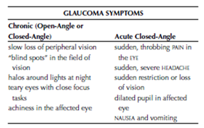
The key symptom of chronic glaucoma, open- angle or closed-angle, is the gradual and painless loss of VISUAL ACUITY and VISUAL FIELD. Often the pattern of progression begins with loss of peripheral (outside) vision. Over time the field of vision becomes increasingly narrow, which people often describe as “tunnel vision.” Other symptoms include excessive tearing (especially with close focus tasks such as reading), halos around lights at night, aching eyes, and headaches. Sudden throbbing PAIN in the eye, loss of vision, severe HEADACHE, halos around lights, and a dilated pupil in the affected eye are symptoms of acute closed- angle glaucoma.
image

Though eye care practitioners routinely use TONOMETRY to screen for increased intraocular pressure, this test alone is not sufficient to detect glaucoma. Detecting glaucoma requires a full OPHTHALMIC EXAMINATION including fundus examination to assess the condition of the optic disk. The ophthalmologist will also conduct a visual acuity test and a peripheral vision test. Other procedures that can help diagnose glaucoma in its early stages or quantify the extent of damage in moderate to advanced glaucoma are ULTRASOUND of the eye and OPTICAL COHERENCE TOMOGRAPHY (OCT).

Treatment Options and Outlook

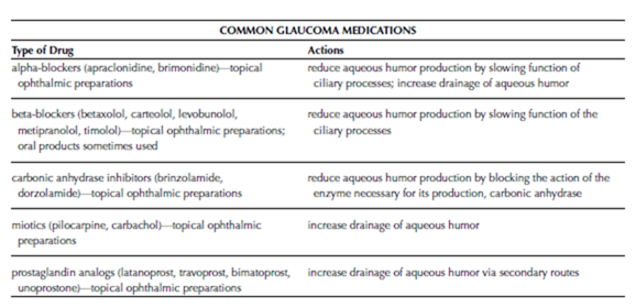
Acute closed-angle requires emergency measures to relieve intraocular fluid and the accumulation of aqueous humor. Such measures typically include a combination of procedures to open the drainage angle, ophthalmic medications to lower intraocular pressure, and systemic medications to draw fluid from cells (osmotics). The ophthalmologist is also likely to administer medications for pain and to minimize NAUSEA and vomiting. Ongoing treatment with glaucoma medications or glaucoma surgery is then necessary. Ophthalmic medications (drops, inserts, and ointments) to open the drainage angle and lower intraocular pressure are the standards of treatment for chronic glaucoma of either form, and typically can control glaucoma for many years.

Surgery becomes an option to treat glaucoma that becomes advanced or does not respond to
image

medication therapy. Surgical treatments for glaucoma include the following:

• For iridotomy, the ophthalmologist uses an ophthalmologic laser to place a small opening in the iris. The opening provides another route of drainage for the aqueous humor and helps keep the iris from blocking the drainage angle in open-angle glaucoma.

• For trabeculoplasty, the ophthalmologist uses an ophthalmologic laser to make numerous “dots” in the trabecular meshwork—the fanlike network of tiny channels at the end of the angle that disperse the draining aqueous humor—to expand its the draining capacity.

• For trabeculotomy, the ophthalmologist places an aqueous shunt, a tiny opening from the anterior chamber through the sclera, to allow aqueous humor to drain to the outside of the eye.

• For cytophotocoagulation, the ophthalmologist uses an ophthalmologic laser to destroy portions of the ciliary processes to reduce aqueous humor production.

Early diagnosis and treatment offer the best opportunity for minimizing vision loss. It is important to diligently follow the directions for using glaucoma medications, as glaucoma requires consistent control. Appropriate treatment can slow the progression of vision loss in most people who have glaucoma.

Risk Factors and Preventive Measures

Age is the most significant risk factor for glaucoma; glaucoma is uncommon in people under age 40 and about two thirds of people who develop glaucoma are over age 65. Glaucoma is more common in people of African American and Asian ethnicity and tends to run in families. Glaucoma also is more likely to develop in people who have hypertension, ATHEROSCLEROSIS, diabetes, and severe MYOPIA (nearsightedness) and in people who take CORTICOSTEROID MEDICATIONS. Prevention focuses on regular ophthalmic examinations to detect glaucoma early in its course.

See also AGE-RELATED MACULAR DEGENERATION (ARMD); CATARACT; LASER SURGERY; VISION HEALTH.

Graves’s ophthalmopathy Changes in the EYE that occur as a result of Graves’s disease, a form of HYPERTHYROIDISM, and occasionally as a result of other forms of thyroid disease. The most prominent feature of Graves’s ophthalmopathy is EXOPH- THALMOS, an outward bulging or protrusion of the eyes that often is the first indication of Graves’s disease. The exophthalmos results from enlarged extraocular muscles (the muscles that move the eye) and edema (swelling due to retained fluid) in the tissues around the eye and within the ocular orbit (eye socket). This circumstance restricts the ability to move the eyes, particularly upward and side to side, as well as to close the eyelids. Graves’s ophthalmopathy can involve only one eye (unilateral) though most often involves both eyes (bilateral). Symptoms and consequences range from mild to severe, with about 5 percent of people experiencing substantial loss of vision that may include loss of the eye. Graves’s ophthalmopathy can appear before there are any indications of Graves’s disease, at the onset of hyperthyroid symptoms, or months to years after the diagnosis of Graves’s disease.

Graves’s ophthalmopathy presents a significant threat to vision. The swelling in and around the orbit pressures the structures of the eye and can compress the OPTIC NERVE, which can result in OPTIC NERVE ATROPHY (the death of cells in the optic NERVE) and permanent VISION IMPAIRMENT. The external pressure against the eye also raises the pressure inside the eye (INTRAOCULAR PRESSURE), which can result in GLAUCOMA. The combination of exophthalmos and restricted lid movement pre- vents the eyelids from closing completely, which allows the CORNEA to become dry. The resulting irritation and INFLAMMATION (KERATITIS) reduces VISUAL ACUITY and also exposes the inner eye to INFECTION. Though the symptoms that threaten vision eventually subside, many of the changes that result, including exophthalmos and vision impairment, are permanent.

Symptoms and Diagnostic Path

The symptoms of Graves’s ophthalmopathy include

• exophthalmos (sometimes called poptosis)

• DIPLOPIA (double vision)

• CONJUNCTIVITIS (inflammation of the inner eye- lids)

• diminished visual acuity (blurry or distorted vision)

• PHOTOPHOBIA (sensitivity to light)

• excessive tearing

As these symptoms are distinctive for Graves’s ophthalmopathy, the doctor often can make the diagnosis based on their presentation. Tests of thyroid HORMONE levels in the BLOOD confirm Graves’s disease, if not already diagnosed. Conventional OPHTHALMIC EXAMINATION and SLIT LAMP EXAMINATION allow the ophthalmologist to assess the status of vision and health of the structures of the eye. A COMPUTED TOMOGRAPHY (CT) SCAN helps assess the extent of orbital swelling and compression of the optic nerve.

Treatment Options and Outlook

The course of Graves’s ophthalmopathy seems to unfold in two stages, regardless of treatment for or status of the underlying hyperthyroidism. The first stage is the acute or active phase, during which symptoms emerge. During this stage, which extends over 18 to 30 months, ophthalmologic treatment focuses on reducing pressure on the eye and stabilizing vision, and may include

• ophthalmic lubricating drops or ointment to keep the cornea hydrated

• patching the eyes at night to protect the corneas during sleep

• NONSTEROIDAL ANTI-INFLAMMATORY DRUGS (NSAIDS) to reduce inflammation and relieve discomfort

• CORTICOSTEROID MEDICATIONS to suppress the body’s immune response

• ANTIBIOTIC MEDICATIONS to treat bacterial infection of the eyelids (BLEPHARITIS), conjunctiva (conjunctivitis), and cornea (keratitis)

In the second stage of Graves’s ophthalmopathy, the progression of symptoms ends. However, the changes that have occurred are permanent. Fibrous deposits replace lymphocytes in the eye muscles, maintaining their enlargement and continuing the exophthalmos. Treatment in this stage targets minimizing these permanent consequences through surgeries to relieve the pressure within the orbit (orbital decompression), reduce the size of the extraocular muscles (myectomy), and reconstruct the eyelids so they close completely over the eye (BLEPHAROPLASTY). Corneal reshaping (keratoplasty) or CORNEAL TRANSPLANTATION may be necessary to restore vision when damage to the cornea is extensive. If infection resulted in loss of the eye, the ophthalmologist will place a PROS- THETIC EYE.

Risk Factors and Preventive Measures Graves’s ophthalmopathy occurs only in conjunction with thyroid disorders, nearly always hyperthyroidism. It may appear months to several years before other clinical indications of hyperthyroidism, or a comparable time after beginning treatment for hyperthyroidism. Prompt diagnosis and treatment are essential to preserve vision, as the changes that occur with Graves’s ophthalmopathy are generally permanent. An ophthalmologist should evaluate any changes in the appearance of the eyes. Regular eye examinations help screen for Graves’s ophthalmopathy as well as other eye health problems.

See also AUTOIMMUNE DISORDERS; BACTERIA; VISION HEALTH.